Confocal Microscopy
Confocal microscopy is able to simultaneously observe the interactions of biological objects with controlled surfaces and the structuring of these surfaces up to the sub-micron level.
Leader
Petithory Tatiana
Contact : tatiana.petithory@uha.fr
Description
Areas of activity :
Confocal microscopy observes and characterizes the interaction of biological objects with controlled surfaces, it also allows topographic characterization at micron and sub-micron level. This in particular leads on to studies in the cellular behaviour in the vicinity of physically or chemically structured surfaces.
Main equipment (strengths of the Institute) :
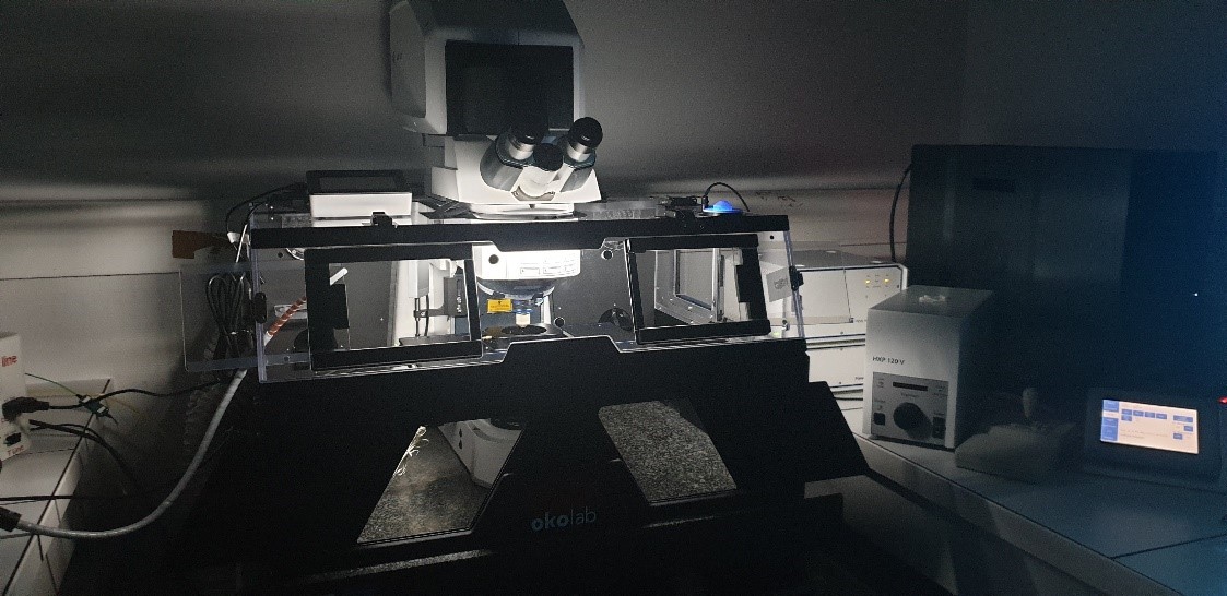
- LSM 800 (Laser Scanning Microscope, ZEISS) Upright configuration equipped with Airyscan® super resolution module and reflexion module (topography)
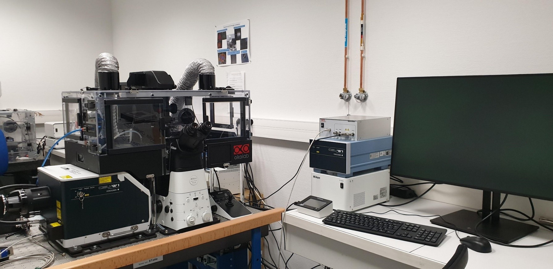
- SD Spinning Disk Confocal Microscope (Yokogawa, Nikon) inverted configuration

Technical description
- LSM 800 (Laser Scanning Microscope, ZEISS) Upright configuration equipped with Airyscan® super resolution module and reflexion module (topography)
|
Statif |
Axio imager M2 Upright |
||||||||||||||||||||||||
|
Motorized stage XY |
• XY Piezo |
||||||||||||||||||||||||
|
LASER diodes |
405nm, 488nm 555nm 640nm |
||||||||||||||||||||||||
|
Detectors |
2 PMTs |
||||||||||||||||||||||||
|
Super Resolution |
AiryScan detector GaAsp |
||||||||||||||||||||||||
|
Objectives |
|
||||||||||||||||||||||||
|
Environnement |
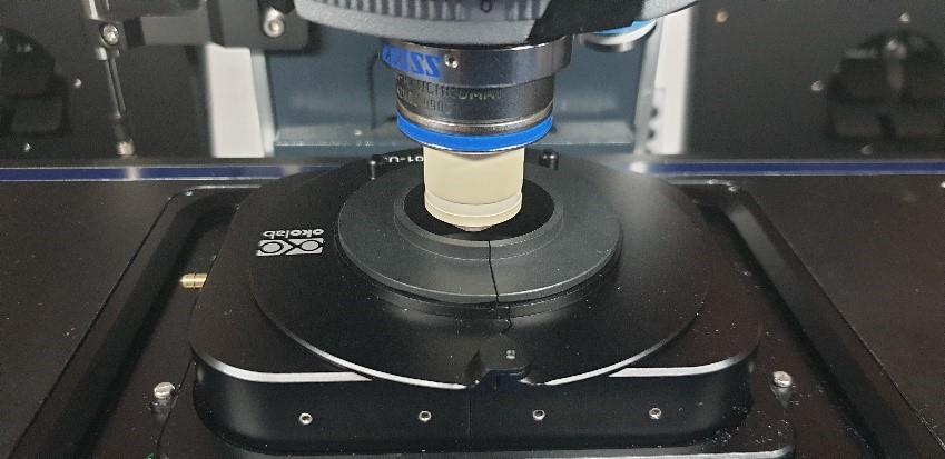
Incubation chamber OKOLAB T° CO2 and Humidity controller |
||||||||||||||||||||||||
|
Analyse |
Software ZEN 2012, IMARIS, Image J ou ICY. |
||||||||||||||||||||||||
|
Accessibility |
Technical training to use the system |
- SD Spinning Disk Confocal Microscope (Yokogawa, Nikon) inverted configuration
|
Statif |
Eclipse Ti2- with « perfect focus » |
|
Motorised stage |
10 nm res/300nm repetition en XY 0,2/0,4 nm/100nm repetition en Z (piezzo) |
|
Light sources |
LUMENCOR SOLA SMII : wavelength 405nm ; 488nm ; 561nm ; 640nm |
|
Objectives |
– 10x oil, water, glycerol ON 0,5 CFI Plan Apochromat – 20x water ON 0,95 CFI APO LWD (0,99-0,90) Lambda S – 60x oil ON 1,4 CFI Plan APO LBDA |
|
SD CSU-W1 |
– Mono- disk (50µm) – Maximal speed 4000 rpm – Field of view 10x16 mm |
|
Laser |
– Oxxius wave length 405-488-561-638 nm |
|
Camera |
– sCMOS Hamamatsu Orca Flash 4.0 – 4,2 MegaPixel – Pixel size 6,2x6,2 µm – EQ 82 % – Air cooling |
|
Incubation |
Incubation chamber OKOLAB T° CO2 and Humidity controller |
|
Software |
– NIS-Element |
IS2M
Bâtiment CNRS
15, rue Jean Starcky - BP 2488
68057 Mulhouse cedex
Bâtiment IRJBD
3 bis, rue Alfred Werner
68093 Mulhouse cedex
tel: (+33)3 89 60 87 00
fax: (+33)3 89 60 87 99
