X-Ray Diffraction (XRD)
X-ray diffraction gives information on the ordered solid at ambient temperature and during heat treatment. The geometric arrangement of the atoms and the distance between them constitute a unique identity card for each compound. X-ray diffusion at small angles studies the organisation of matter over a few nanometers.
.
Leaders
Laure Michelin and Ludovic Josien
Contacts : laure.michelin@uha.fr or ludovic.josien@uha.fr
Description
Areas of activity :
The activities of the platform focus on the structural characterisation of all types of solid materials by X-ray diffraction or diffusion : identification of phases, study of crystallinity or organisation, reflectometry, grazing incidence, structural determination on powder, observation of phase transitions in temperature and controlled atmosphere. In addition, these characterisations can be supplemented by X-ray Fluorescence spectrometry analyses which make it possible to determine the chemical composition of a sample and thus facilitate structural identification.
Main equipment (strengths of the Institute) :
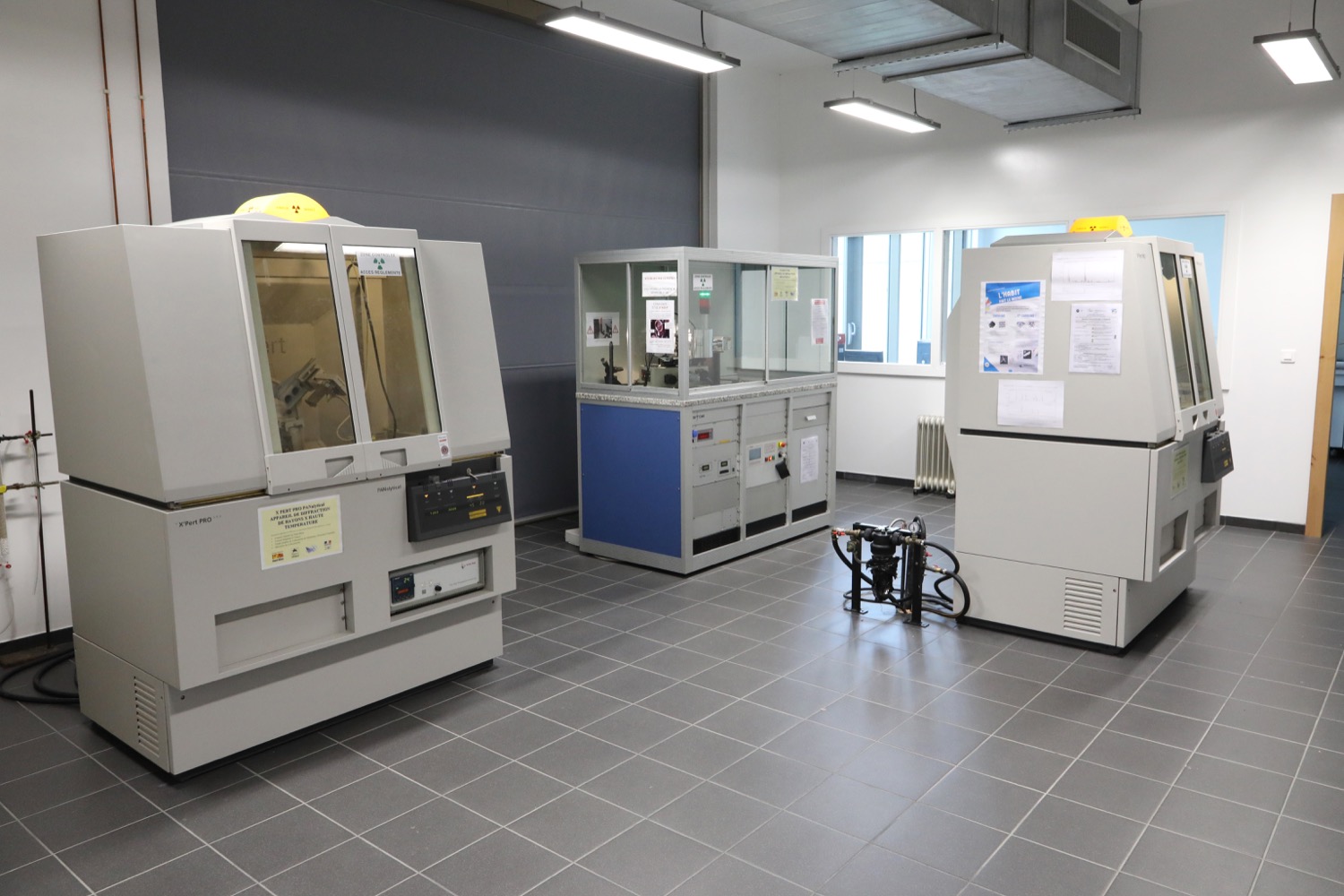
The platform has 3 diffractometers, one thermodiffractometer, and an X-ray fluorescence spectrometer :
- 3 powder diffractometers : 2 in reflection geometry with autosampler (Bruker D8 ADVANCE and PANalytical, X’Pert PRO MPD) and 1 in transmission geometry (STOE Stadi-P and Bruker D8 ADVANCE)
- 1 thermodiffractometer (PANalytical, X’Pert PRO MPD) equipped with a temperature chamber (Anton Paar HTK1200)
- 1 Wavelength Dispersion X-Ray Fluorescence Spectrometer (PANalytical, Zetium)
Technical description
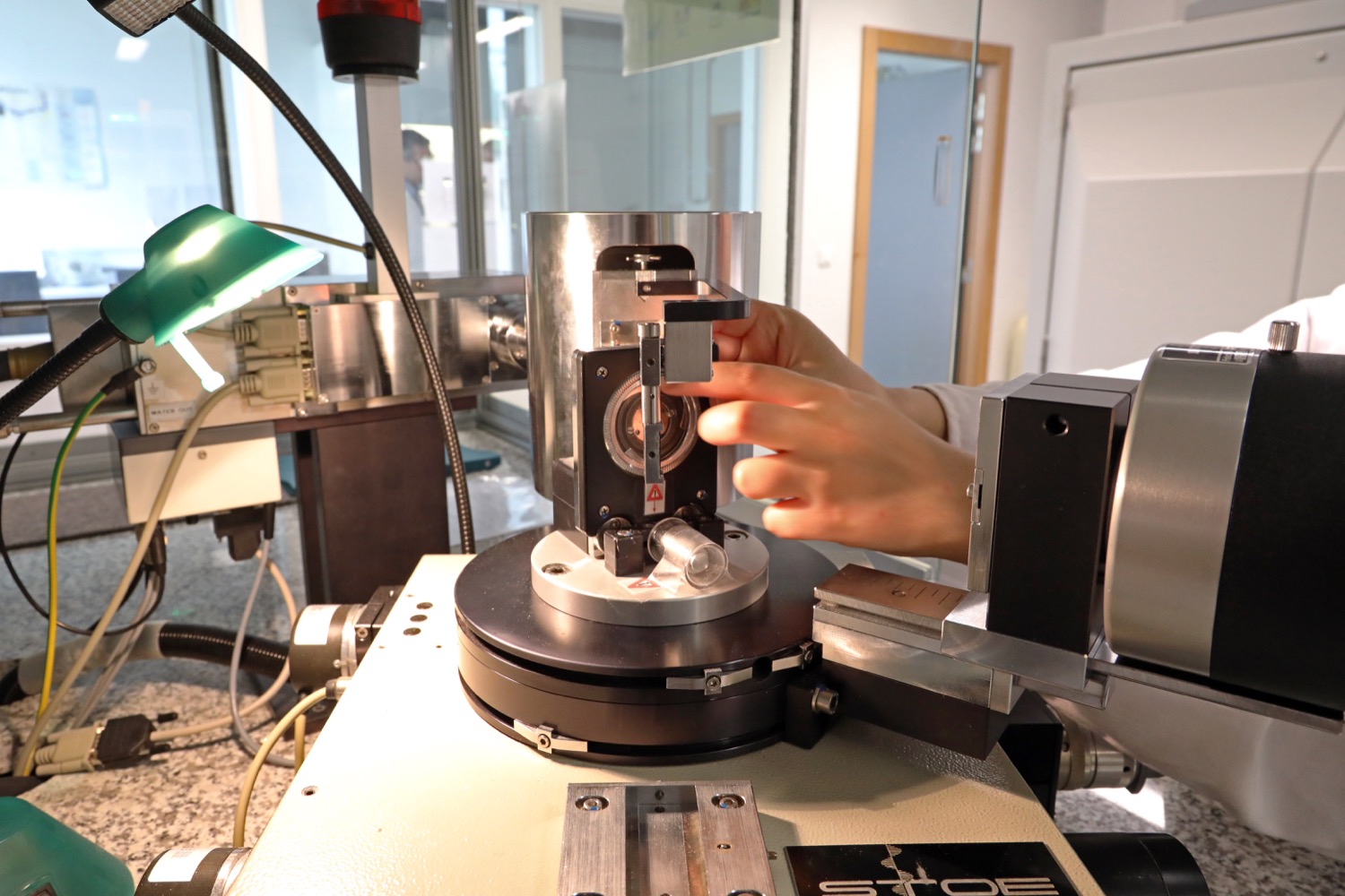
PANalytical X-ray Diffractometer, model X'Pert PRO MPD with feeder
| Location | rue A. Werner |
| Year of commissioning | 2004 |
| Anticathode | copper (λ = 1.54Å) |
| Geometry | reflection (Bragg-Brentano) |
| Goniometer | vertical, system θ – θ |
| Angular domain of analysis | 0.5 to 150° 2θ |
| Minimum angular pitch | 0.002° 2θ |
| Type of detector | PIXcel 1D |
| Monochromator | carbon graphite (002) – rear position – removable |
| Passing capacity | 45 samples |
| Sample preparation constraint | powder samples or solid samples (diameter < 43mm, height < 6mm) |
| Self-service accessibility | no – Samples are prepared by the users but the autosampler is programmed and checked by the Technical Authority |
Contact : Laure Michelin, Ludovic Josien
PANalytical X-Ray Thermodiffractometer, X'Pert PRO MPD model
| Location | rue A. Werner |
| Year of commissioning | 2003 |
| Anticathode | copper (λ = 1.54Å) or chromium (λ = 2.29Å) |
| Geometry | reflection (Bragg-Brentano) or transmission (Debye-Scherrer) |
| Goniometer | vertical, system θ – 2θ |
| Angular domain of analysis | 0.5 to 130° 2θ |
| Minimum angular pitch | 0.002° 2θ |
| Type of detector | X’Celerator or proportional detector |
| Monochromator | Germanium (111) – front position – removable |
| Specific configuration | Anton Paar HTK1200 high temperature chamber under a specific atmosphere (neutral gas or oxygen, no reducing gas) and controlled heating up to 1200°C (manufacturer’s data) |
| Sample preparation constraint | powdered samples. Possibility of solid samples for some configurations (height < 6mm) |
| Self-service accessibility | partial yes for analysis after training by the technical manager no for configuration changes. |
Contact : Laure Michelin, Ludovic Josien
Bruker D8 ADVANCE A25 X-ray diffractometer
| Location | rue J. Starcky |
| Year of commissioning | 2016 |
| Anticathode | copper (λ = 1.54Å) |
| Geometry | reflection (Bragg-Brentano), transmission |
| Goniometer | vertical, scanning θ – θ |
| Angular domain of analysis | Variable according to accessory and type of acquisition, typically 0.5 ° to 130° 2θ (reflection) |
| Minimum angular pitch | 0.0001° 2θ |
| Autochanger capacity (autosampler) | 15 |
| Detector | LynxEye XE-T with power discrimination (<380eV in high resolution mode) |
| Monochromator | No |
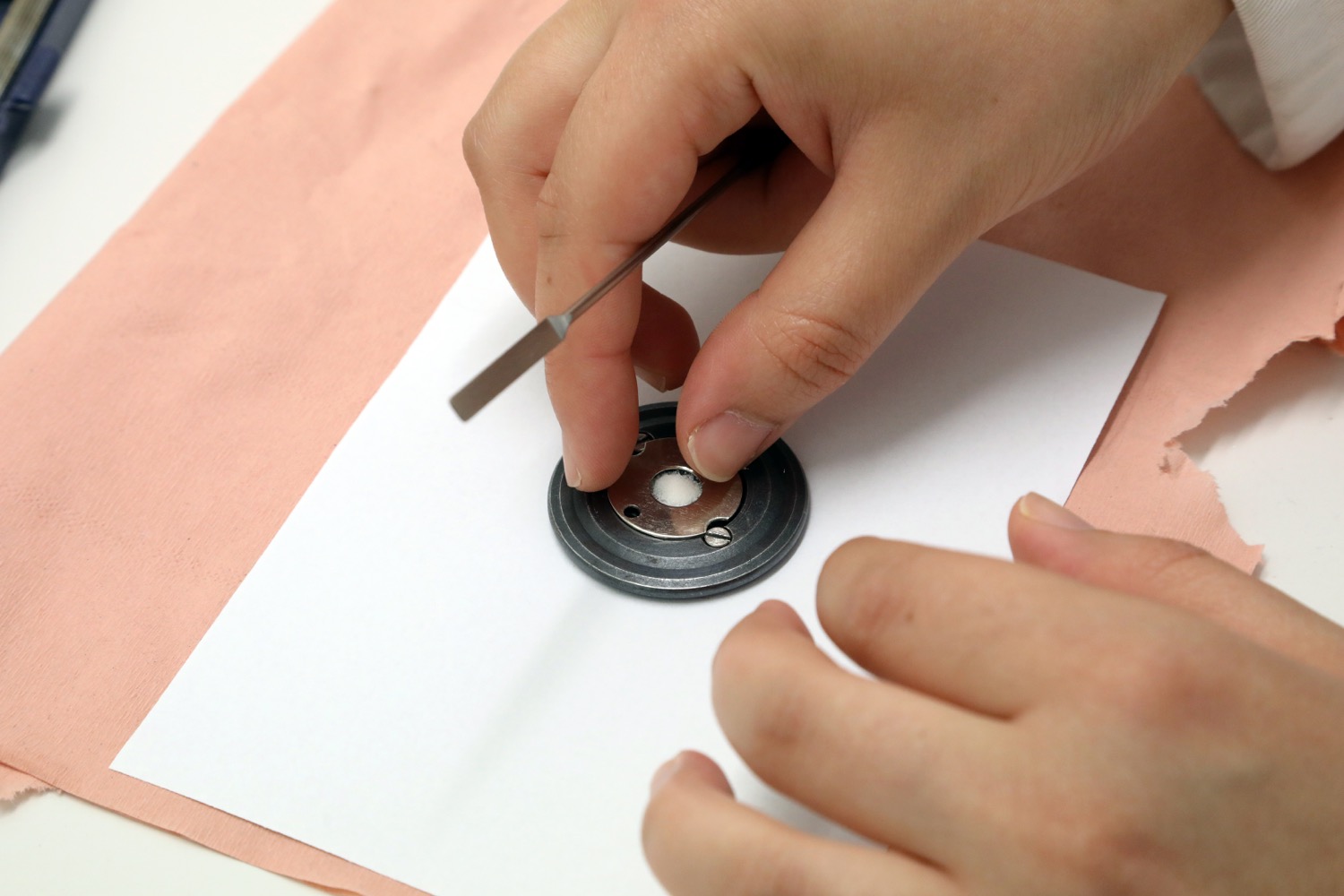
| Sample preparation constraint | Powder samples (400mg), fibres or solid samples with conditions. |
| Accessories | Compact and motorized XYZ stage allows grazing incidence or reflectivity measurements to be taken. |
| Self-service accessibility | No. The samples are prepared by the users but the Autochanger is programmed and verified by the person in charge of the machine. |
Contact : Jean-Marc Le Meins, Simon Gree
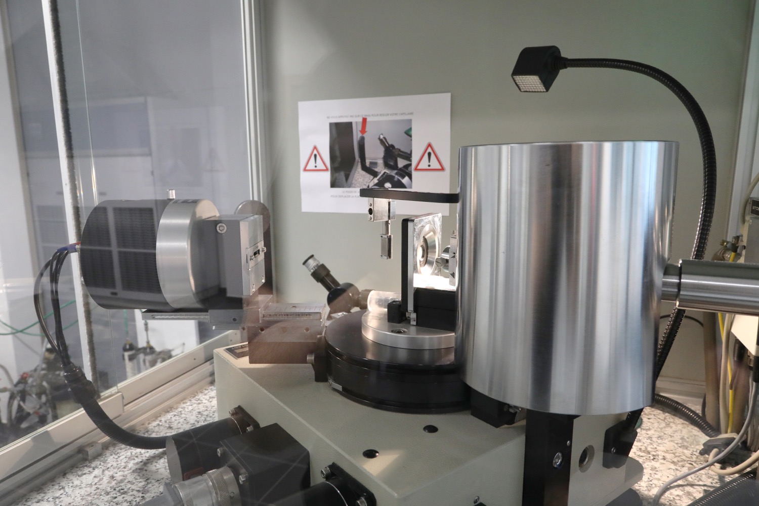
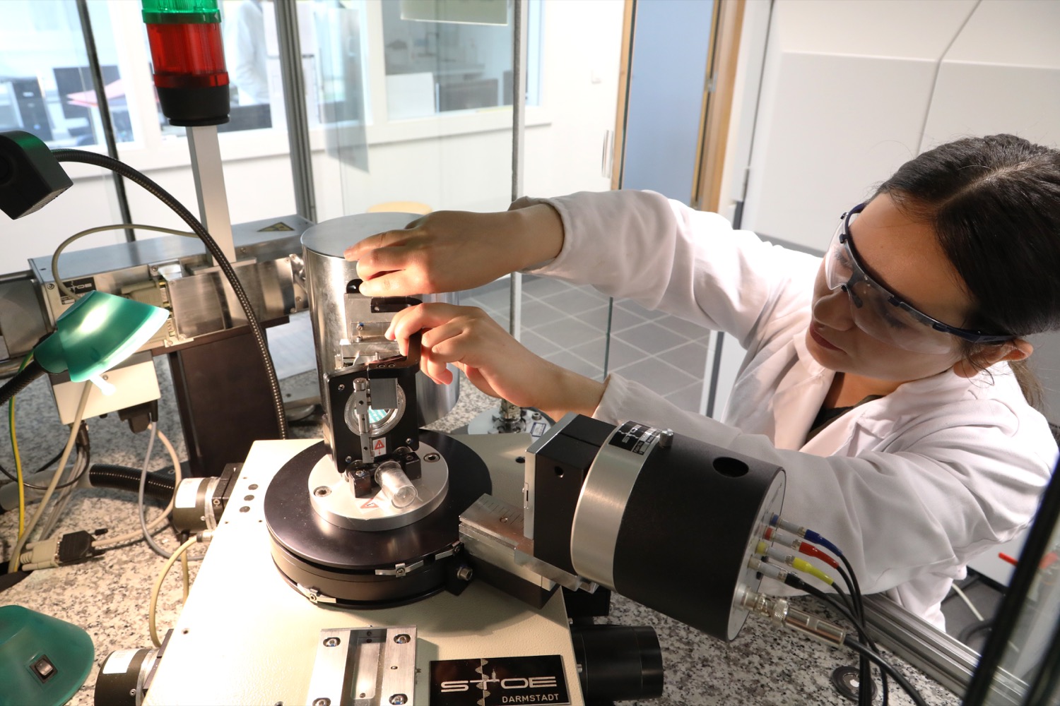
STOE X-ray diffractometer, STADI-P model
| Location | rue A. Werner |
| Year of commissioning | 1994 |
| Anticathode | copper (λ = 1.54Å) |
| Geometry | transmission (capillary or flat plate) |
| Goniometer | horizontal, scanning θ -2θ |
| Angular domain of analysis | 3.0 to 110° 2θ |
| Minimum angular pitch | 0.01° 2θ |
| Detector | linear with spatial location (Position Sensitive Detector) |
| Monochromator | in germanium (111), forward position – fixed |
| Sample preparation constraint | powder samples only |
| Self-service accessibility | yes – after training by the technical manager. |
Contact : Laure Michelin, Ludovic Josien
PANalytical Wavelength Dispersion X-Ray fluorescence Spectrometer, Zetium model
| Location | rue A. Werner |
| Year of commissioning | 2017 |
| Anticathode | Rhodium 4kW |
| Detector | Scintillator, Gaseous and ED Flux (for Small Spot Mapping) |
| Domain of analysis | Uranium Sodium Analysis + fluorine boron detection |
| Specific configuration | Option to carry out an elementary mapping of the sample |
| Sample preparation constraint | powdered samples or solid samples (diameter less than 40 mm) |
| Self-service accessibility | no – Samples are prepared by the users but the autosampler is programmed and checked by the technical manager. |
Contact : Laure Michelin, Ludovic Josien
IS2M
Bâtiment CNRS
15, rue Jean Starcky - BP 2488
68057 Mulhouse cedex
Bâtiment IRJBD
3 bis, rue Alfred Werner
68093 Mulhouse cedex
tel: (+33)3 89 60 87 00
fax: (+33)3 89 60 87 99
