Electronic microscopy
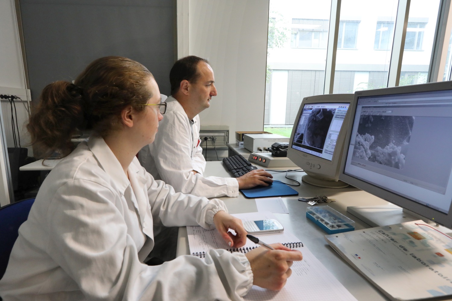
Electron microscopy makes it possible to observe all types of materials from the nano scale to the macro scale. The information obtained provides information on morphology and surface chemistry as well as internal organisation and chemical composition.
Leader
Vidal Loïc
Contacts: loic.vidal@uha.fr ou ludovic.josien@uha.fr
Description
Areas of activity:
The equipment of the platform (scanning electron microscopes and transmission electron microscopes) makes it possible to carry out morphological, chemical and crystallographic characterisations of all types of materials (polymers, porous solids, carbons, biological objects, etc.).
Main equipment (strengths of the Institute):
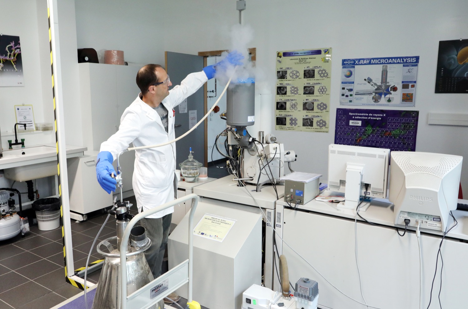
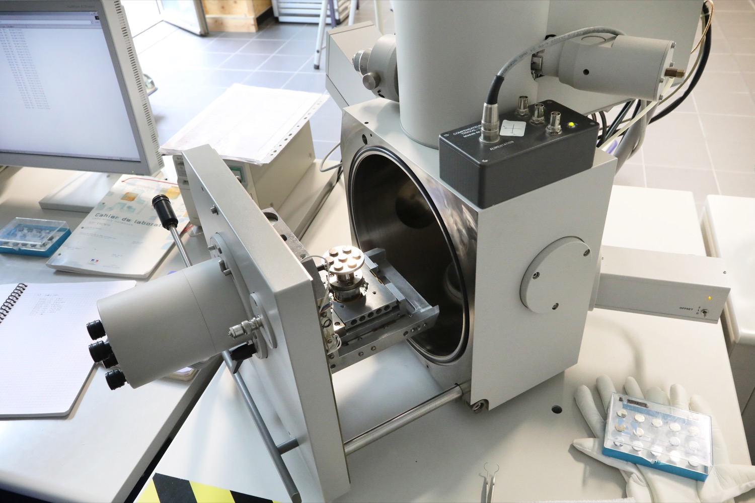
The platform is equipped with 5 microscopes, a ion slicer and an ultramicrotome for the preparation of samples:
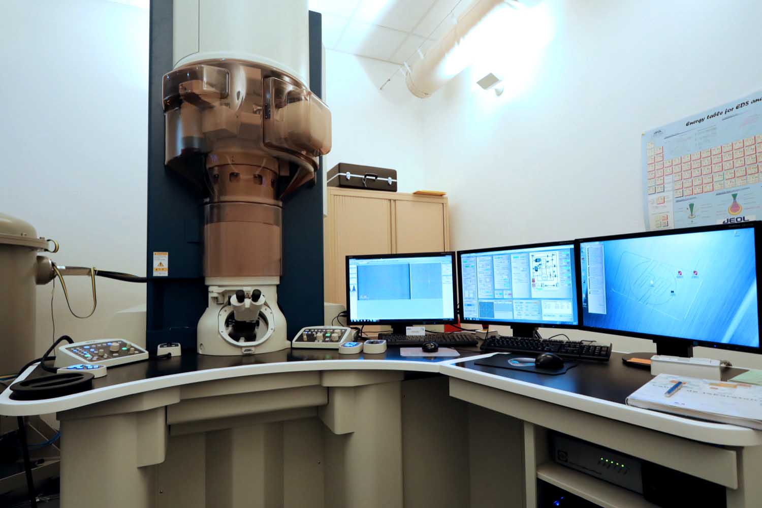
- 1 atomic-resolution transmission electron microscope (JEOL, ARM-200F) equipped with a chemical analysis system
- 1 transmission electron microscope (Philips, CM200)
- 1 scanning electron microscope (Philips, XL30) equipped with a Kammrath liquid cryostage and 4 Imina nanoprobing MiBots
- 1 high resolution scanning electron microscope (Jeol, JSM-7900F) equiped with a chemical analysis system (Bruker, Quantax)
- 1 ion slicer (Jeol EM-09100IS)
- 1 environmental-type scanning electron microscope (FEI, Quanta 400) equipped with a chemical analysis system
- 1 cryogenic ultramicrotome (Leica, EM UC7)
Technical description
Philips Scanning Electron Microscope, XL30 model
| Location | rue A. Werner |
| Canon | field effect – W/ZrO2 point |
| Operating mode | high vacuum |
| Resolution | About 2 nanometers |
| Sample preparation constraint | samples must be conductive (metallization required for insulation samples) |
| Means of control relating to the environment | air conditioner and cold water loop |
| Imina miBots TM | 4 micro robots indepently driving of their own probe (a contacting tip or an optical fiber). The positioning accuracy of a contact tip of a micromanipulator on an object is less than a micrometer. The robots are designed to be able to measure very low currents. |
| Kammrath Cryostage | liquid nitrogen cryostage which allows samples to be cooled down to -195°C. The stage is also equiped with a heater that can reach 300°C. It is possible to use the cryostage and the 4 microrobots together. This coupling allows current measurements to be made at low temperatures |
| Self-service accessibility | yes for IS2M PhD students and students with a large number of samples and after training by the technical manager |
Contact: Ludovic Josien
FEI Environmental Scanning Electron Microscope, Quanta 400 model
| Location | rue J. Starcky |
| Canon | thermo-ionic, filament W |
| Operating mode | high vacuum low vacuum (up to 1 torr of water in the chamber, observation of insulating samples) environmental (up to 20 torr of water in the chamber, observation of hydrated samples) |
| resolutions | high vacuum: About 5 to 10 nanometres low vacuum: About 10 to 20 nanometres environmental: > at 20 nanometres |
| Sample preparation constraint | high vacuum observations: samples must be conductive (metallization required for insulation samples) observations in low vacuum and environmental: no prior preparation even for insulation samples |
| Accessories for use in low vacuum and environmental | Peltier plate: range of accessible temperatures: -5 to 40°C warming plate: range of accessible temperatures: 20 to 1000°C traction plate for polymer films: range of stretches: 0 to 200% |
| EDX | all detectable and quantifiable elements from boron |
| Means of control relating to the environment | air-conditioner |
| Self-service accessibility | no |
Contact: Loic Vidal
Transmission electron microscope Philips, CM200
| Location | rue J. Starcky |
| Canon | thermo-ionic, LaB6 filament |
| Operating mode | high vacuum |
| Resolution | 0.3 nanometre |
| Sample preparation constraint | the samples must have a thickness of less than 100 nm (obtained by grinding or ultramicrotomy) |
| EDX | all detectable and quantifiable elements from boron |
| Electronic diffraction | possibility of determining inter-reticular distances in the case of crystallized solids |
| Means of control relating to the environment | air conditioner and cold water loop |
| Self-service accessibility | no |
Contact: Loic Vidal
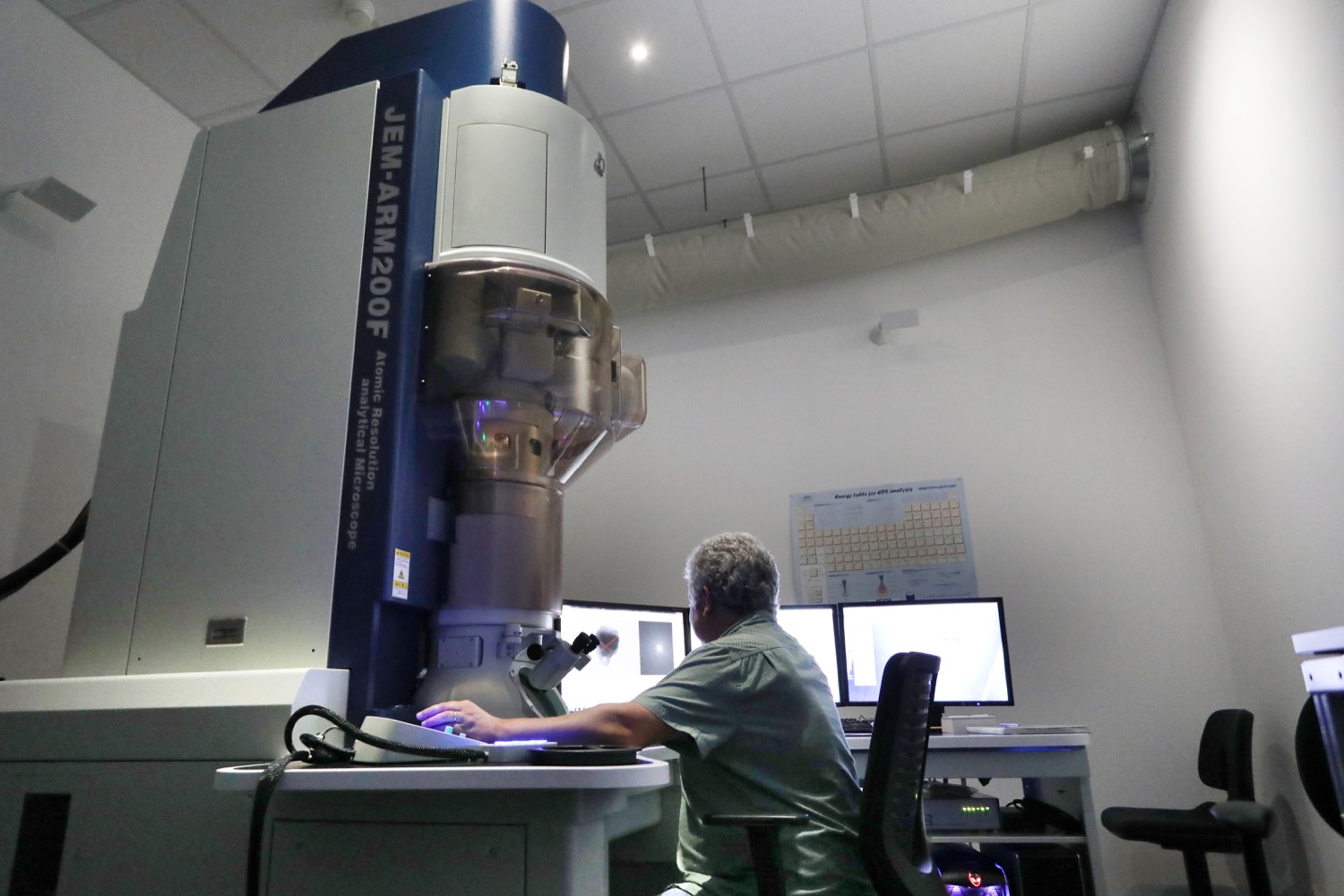
Transmission electronic microscope JEOL, ARM200
| Location | rue J. Starcky |
| Canon | cold FEG |
| Operating mode | high vacuum |
| Configuration | TEM, STEM: at 80 or 200kV Presence of an objective lens corrector |
| Resolution | 80pm (TEM in network border mode) |
| Sample preparation constraint | the samples must have a thickness of less than 100 nm (obtained by grinding or ultramicrotomy) |
| EDX | semi-quantitative analyses: all detectable and quantifiable elements from boron. Chemical mapping |
| Electronic diffraction | possibility of determining inter-reticular distances in the case of crystallized solids |
| Tomography | 3D reconstruction of objects observed |
| Means of control relating to the environment | air conditioner and cold water loop |
| Self-service accessibility | no |
Contact: Loic Vidal
JEOL High Resolution Scanning Electron Microscope, JSM-7900F model
| Location | rue A. Werner |
| Gun | Schottky field emission gun |
| Operating mode | High vaccum with chamber SE and BSE detectors + In-lens electrons detectors + transmission electrons detectors (BF, DF, HAADF)
Low vaccum with chamber SE and BSE detectors for the observation of insulating samples (maximal chamber pressure : 300 Pa of N2 ) |
| Resolutions | High vacuum : 0.5 nanometers Low vacuum : 1.4 to 4 nanometers depending on the pressure value |
| Sample preparation constraint | – General case : maximum sample size admitted 5 cm in diameter and 4 cm in height – Transmission mode : maximum sample size admitted 3 mm in diameter and 100 nm in height |
| Accessories | – Plasma cleaner available in the exchange chamber |
| EDX | Two 30mm² silicon drift detector All elements detectable from boron and quantifiable from fluorine |
| Means of control relating to the environmen | air conditioner and cold water loop |
| Self-service accessibility | no |
Contact: Ludovic Josien
IS2M
Bâtiment CNRS
15, rue Jean Starcky - BP 2488
68057 Mulhouse cedex
Bâtiment IRJBD
3 bis, rue Alfred Werner
68093 Mulhouse cedex
tel: (+33)3 89 60 87 00
fax: (+33)3 89 60 87 99
